Scientific Exhibits
Home


Return to the top of
Section VII


View Previous Case

View Next Case


DISTANT 
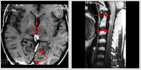
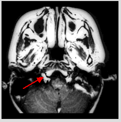
METASTASIS - The Hypoglossal Nerve

Now viewing: Case 12

Case 12 - Recurrent mixed germ cell tumor of pineal area with leptomeningeal spread and perineural metastasis to the hypoglossal nerve

This 14-year-old boy had mixed germ cell tumor of the pineal area and was treated with surgery, radiation therapy and chemotherapy elsewhere. Admission MRI showed recurrent tumor of the pineal area with heptomeningeal seeding and perineural metastasis to hypoglossal nerve. Patient was then treated conservatively.
case_12.gif (29708 bytes)
Axial post-contrast T1-weighted MR image (TR 440/TE 26) of the brain at the level of the third ventricle: contrast-enhanced tumor mass in the pineal region (1) and heptomeningeal seeding in the straight sinus and falx cerebri (2). Midline sagittal post-contrast T1-weighted MR image (TR 414/TE 30): nodular and linear contrast-enhanced leptomeningeal seeding within the cervical spinal canal (arrows).

case_12a.gif (15289 bytes)

Axial post-contrast T1-weighted MR image (TR 440/TE 26) at the level of the clivus: contrast-enhanced enlarged hypoglossal nerve with expansion of the hypoglossal canal (arrow).


©2002 The Levit Radiologic - Pathologic Institute
1100 Holcombe Blvd, Houston, TX 77030
 (USA) / 713-792-2728     

Last updated; February 2002 - contact  Webmaster 

©2002 The University of Texas M. D. Anderson Cancer Center
1515 Holcombe Blvd, Houston, TX 77030
1-800-392-1611 (USA) / 1-713-792-6161     Legal Statements