|

|
REGIONAL
EXTENSION
Now viewing: Case
14
Case 14 - Squamous cell
carcinoma of right external auditory canal with possible perineural
extension to mastoid segment of facial nerve
| A
49-year-old male had tumor mass in the right external auditory
canal. Biopsy revealed squamous cell carcinoma. Resection of the
tumor mass in the external auditory canal was performed with
removal of the superficial limits of the external auditory bony
canal. Histopathology revealed invasive squamous cell carcinoma
of the right external auditory canal with focal squamous cell
carcinoma of the overlying tissue of the mastoid. The vascular,
lymphatic and perineural invasion were not seen, however, the
actual tumor volume is small and thus may not be representative. |
|

|
|
|
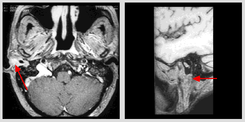
| Axial
post-contrast T1-weighted MR image (TR 600/TE 12) at the
level of auditory canal: A contrast-enhanced soft tissue
mass in the right external auditory canal (arrow). |
|
Sagittal
post-contrast T1-weighted MR image (TR 600/TE 12) 40 mm to
the right: the mastoid segment of the facial nerve is well
shown with minimal enhancement suggesting early tumor
involvement.
|
|
|
|
©2002
The Levit Radiologic - Pathologic Institute
1100 Holcombe Blvd, Houston, TX 77030
(USA) / 713-792-2728
Last updated;
February 2002 - contact Webmaster
|
©2002
The University of Texas M. D. Anderson Cancer Center
1515 Holcombe Blvd, Houston, TX 77030
1-800-392-1611 (USA) / 1-713-792-6161 Legal
Statements
|
|
|
|


