Scientific Exhibits
Home


Return to the top of
Section VII


View Previous Case

View Next Case


DISTANT METASTASIS - The Trigeminal Nerve

Now viewing: Case 7

Case 7 - Melanoma of right cheek with metastasis to maxillary division

This 79-year-old female had melanoma of right cheek with perineural metastasis to the second division of the right trigeminal nerve. Partial maxillectomy was performed with excision of soft tissue melanoma. Histopathology revealed melanoma in the soft tissue overlying anterior wall of the right maxilla and perineural invasion of the right infraorbital nerve with tumor present at margins of resection.
case_07.gif (23241 bytes)
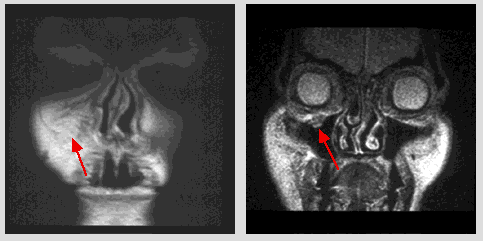
Coronal post-contrast T1-weighted MR image (TR 550/TE 14) at the level of the anterior wall of the maxillary sinus: a contrast-enhanced soft tissue tumor mass within the marked soft tissue thickening anterior to the right maxillary sinus. Coronal T2-weighted MR image (TR 2000/TE 80) at the level of the anterior maxillary sinus: enlargement of the right infraorbital nerve with expansion of the canal is well shown (arrow).
case_07a.gif (24595 bytes)
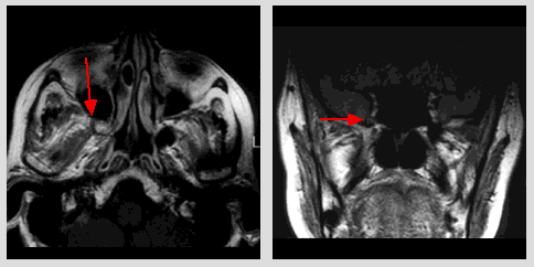
Axial T1-weighted MR image (TR 550/TE 14) at the level of the nasopharynx and maxillary sinus: soft tissue mass with low signal intensity in the right pterygopalatine fossa with destruction of the bony boundaries.


Coronal PD-weighted MR image TR 2000/TE 18) at the anterior sphenoid sinus: slight enlargement of the right foramen rotundum with an abnormal signal intensity (arrow).

©2002 The Levit Radiologic - Pathologic Institute
1100 Holcombe Blvd, Houston, TX 77030
 (USA) / 713-792-2728     

Last updated; February 2002 - contact  Webmaster 

©2002 The University of Texas M. D. Anderson Cancer Center
1515 Holcombe Blvd, Houston, TX 77030
1-800-392-1611 (USA) / 1-713-792-6161     Legal Statements