Scientific Exhibits
Home


Return to the top of
Section IV 


View Next Image


NORMAL DIAGNOSTIC IMAGING OF TRIGEMINAL, FACIAL AND HYPOGLOSSAL CRANIAL NERVES

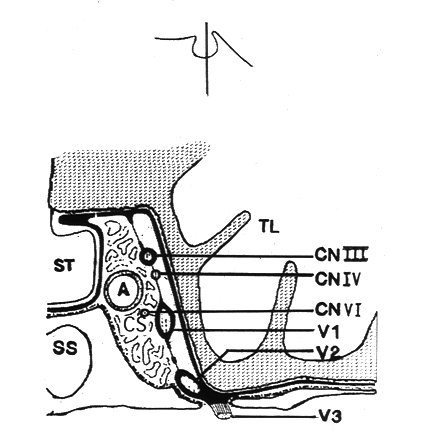
TRIGEMINAL CRANIAL NERVES  Now viewing : Figure 6


Figure 6 - Schematic coronal section through the middle part of the sella turcica showing the proximal branches of the trigeminal nerve within the cavernous sinus

cavernous_sinus.gif (41486 bytes)
Label Key
A - Internal carotid artery
CS - Cavernous sinus
TL - Temporal lobe
ST - Sella turcica  
SS - Sphenoid sinus
CNIII - Oculomotor nerve 
CNVI - Abducens nerve
CNIV - Trochlear nerve
CNV1 - Ophthalmic division of
               the trigeminal nerve 
CNV2 - Maxillary division of the
               trigeminal nerve
CNV3 - Mandibular division of
              the trigeminal nerve


©2002 The Levit Radiologic - Pathologic Institute
1100 Holcombe Blvd, Houston, TX 77030
 (USA) / 713-792-2728     

Last updated; February 2002 - contact  Webmaster 

©2002 The University of Texas M. D. Anderson Cancer Center
1515 Holcombe Blvd, Houston, TX 77030
1-800-392-1611 (USA) / 1-713-792-6161     Legal Statements