MM Learn
   Scientific Exhibits

   Soft-Tissue
  
Sarcoma Home

     Abstract

     Methods & 
    
Materials

     References

     Quiz

     Help

     Contact


MR IMAGING GUIDELINES AND PROTOCOL

INTRODUCTION 

The goal of the radiologist should be to:

  • Detect Tumor
  • Localize Tumor
  • Determine Extent
  • Characterize Tumor
  • Attempt Diagnosis

In each individual institution standardization of pulse sequences to achieve consistency in scanning is important. The area of interest may be marked on the skin ( eg. by docusate sodium gel capsules [oil-based stool softener]). It is important to mark the superior and inferior aspects of the surgical scar [following excision of soft-tissue sarcoma] to ensure scanning through the entire length of the scar to detect recurrence, if any. Gradient sequences and MR angiography may be utilized. Use of contrast agents in pre treated sarcomas may identify areas of necrosis. Some current research projects in our department include attempts to quantify this necrosis and serially observe increase in necrosis, if any, secondary to chemotherapy and/or radiation therapy. Contrast media administration may help to distinguish benign fluid containing lesions and may also help in the post operative cases to distinguish recurrence from post surgical change.

Images

Click on image(s) to view JPEG image(s).
1.
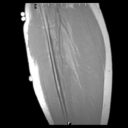
Sagittal T1W MR image which marks the superior and inferior extent of a post-surgical scar utilizing "oil-based" capsules. Such an image can serve as the localizer for axial scans. Note capsules taped on skin serving as markers (arrows on JPEG image).
2.
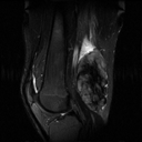
18-year-old male with desmoid fibromatosis in distal thigh. Sagittal fast spin-echo T2W MR image (TR/Effective TE - 9000/85) reveals heterogeneous mass in distal thigh.
3.
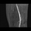
MR Time of flight 2D angiogram (TR/TE flip angle-30/7.7/60°) acquired in the axial plane with reconstruction and display in coronal and sagittal planes shows displaced femoral vessel without encasement.
4.
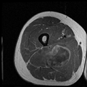
35-year-old male with unclassified myxoid sarcoma of thigh. Gd+DTPA enhanced T1W MR image reveals areas of "necrosis" (arrows on JPEG image) within tumor. Surgery was performed two days following the MR study and pathological examination revealed approximately 50-60% therapy induced necrosis within tumor.


Copyright ©2003 All rights reserved.
The University of Texas M. D. Anderson Cancer Center
1515 Holcombe Blvd, Houston, TX 77030
1-800-392-1611 (USA) / 1-713-792-6161     Legal Statements
Last updated; September 2003


main menu next