

|
|
Cyclin
D1 FISH Test
Detection of Cyclin D1 Translocation
by FISH is Highly Specific and Sensitive in the Diagnosis of Mantle Cell
Lymphoma (MCL)
Ruth L. Katz, M.D., Director
Image Cytometry Diagnostic Laboratory, University of
Texas, M.D. Anderson Cancer Center
Background
-
Mantle cell lymphoma (MCL) is a subtype of non-Hodgkins
lymphoma characterized by poor prognosis and a median survival of approximately
3 years.
-
On the basis of morphology and immunophenotype alone, mantle
cell lymphoma is difficult to distinguish from indolent lymphomas and leukemias.
-
Cytogenetically, a t(11:14)(q13;q32) is associated with 75%
of mantle cell lymphomas.
-
PCR for the major translocation cluster (MTC) region of the
bcl-1
is positive in only 30-40% of cases.
-
Southern blot analysis detects translocations in approximately
50% to 70% of the cases.
-
Translocation breakpoints are scattered within the approximately
120-kb bcl-1 region adjacent to cyclin D1, whereas part of
them are clustered in the MTC.
The translocation leads to overexpression of cyclin
D1 due to juxtaposition of the Ig heavy chain gene enhancer on 14q32 to
the cyclin D1 gene on 11q13.
Cyclin D1/CEP11
-
Cyclin D1 amplification is common in many types of
cancer, second only to the HER2/neu (erb-b2) oncogene.
-
The LSI Cyclin D1 is approximately 300 kb in size.
-
This probe may be used to determine the copy number of the
Cyclin
D1 locus, or as an enumerator probe for chromosome 11 in interphase and
metaphase cells.
Fluorescent
in
situ Hybridization (FISH) Analysis
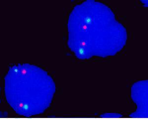 |
Figure 1. Interphase FISH from a non-MCL patient. Cells
showing 2 green signals (Centromere 11) and 2 orange signals (Cyclin D1
at chromosome 11q13). |
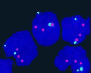 |
Figure 2. Interphase FISH from a MCL patient. Most of
cells showing 2 green signals (Centromere 11) and 3 orange signals (Cyclin
D1 at chromosome 11q13). |
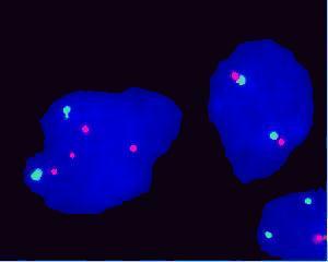 |
Figure 3. Interphase FISH from a blastoid MCL patient.
Cell on the left side showing 2 green signals (Centromere 11) and 4 orange
signals (Cyclin D1 at chromosome 11q13). |
How to request the FISH test
When Mantle Cell Lymphoma is in the differential diagnosis,
you may request this highly sensitive FISH test by calling Research Image
Diagnostic Lab at (713)792-4087 or (713) 792-4088. Fine needle aspiration
samples in culture media or PBS, peripheral blood samples in sodium heparin
vacutainer, or bone marrow samples in sodium heparin vacutainer will be
acceptable for the test. |
|
|