|
|

|
|


|
| |
|

|
|
POST
SURGICAL FLUID COLLECTIONS
|
|
POST OPERATIVE
SEROMA
Patient 2
68-year-old
male who underwent excision for a pleomorphic liposarcoma of the
left thigh at an outside institution.
|
Click
on image(s) to view JPEG image(s).
|
| 1. |
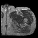
Axial
T2W MR image nine months post surgery reveals postoperative
seroma (arrow on JPEG image). Repeat surgery at our institution
3 months following the MR examination revealed a cystic space
and histological examination revealed focal foreign body giant
cell inflammation in fibroadipose tissue. No residual tumor
was identified.
|
| 2. |
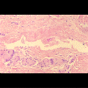
Representative
photomicrograph from 'cystic' space reveals space lined by
denuded epithelium and fibrocollagenous wall with giant cells.
(Original magnification, X120, H-E Stain). Prolonged retention
of some postoperative seromas may be secondary to such spaces
formed after surgery.
|
|
Copyright
©2003 All rights reserved.
The University of Texas M. D. Anderson
Cancer Center
1515 Holcombe Blvd, Houston, TX 77030
1-800-392-1611 (USA) / 1-713-792-6161 Legal
Statements
Last updated; September 2003
|
|
|