|
Click
on image(s) to view JPEG image(s).
|
| 1. |
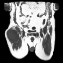
(01/09/91)
Coronal T1W MR image reveals intermediate signal intensity
mass in right thigh. Patient underwent wide local excision.
|
| 2. |
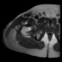
(09/05/91)
Axial T1W MR image obtained 8 months after surgery reveals
low intensity seroma.
|
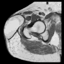
| 3. |

(09/05/91)
Axial T2W MR image reveals a small focus of layering hemorrhage
within the seroma (arrow on JPEG image). Fine needle aspiration
performed the same day revealed serous fluid and no malignancy.
|
| 4. |
(01/11/93)
Axial T1W MR imaging obtained about 16 months following previous
MRI and 2 years after the surgery reveals high signal intensity
fluid indicating that ongoing hemorrhage has occurred. No
history of recent trauma.
|
| 5. |
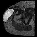
01/11/93)
Axial T2W MR image reveals high intensity hemorrhage and heterogeneous
appearance of fluid collection. Fine needle aspiration at
this time revealed hemorrhagic fluid and no malignancy.
|
