|
|

|
|


|
|
|
|

|
|
MIMICS
OF SOFT-TISSUE SARCOMAIN:
BENIGH LESIONS
|
|
INTRODUCTION
EXTRA-ABDOMINAL
DESMOID TUMOR
- Fibroblasts
- fibrocytes, abundant collagen
- M > F, any
age but more frequent up to 40 years
- Limbs, trunk,
head and neck, shoulder, buttock, posterior thigh, popliteal fossa,
sura, arm, forearm - deep soft tissues
- Grows by
infiltration,
- Grows progressively
but not rapid
- Treatment
surgical
|
Click
on image(s) to view JPEG image(s).
|
| |
Patient
11
30-year-old female with surgically proven aggressive desmoid
fibromatosis in left proximal thigh.
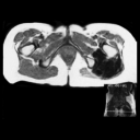
Axial T1W MR image reveals low signal intensity mass in
proximal left thigh. Similar low signal intensity was noted
on T2W MR images. Signal intensity of desmoid tumors varies
with degree of cellularity and amounts of fibrous and collangenous
tissue. High signal on T2W image does not exclude desmoid
fibromatoses.
|
| 1. |
Patient
12
18-year-old male with surgically proven desmoid fibromatosis
in distal thigh.
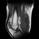
Sagittal T1W MR image reveals intermediate signal intensity
mass in distal thigh with interspersed areas of low signal
intensity.
|
| 2. |
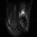
Sagittal FSE T2W MR image reveals heterogenous mass with
central areas of low signal intensity secondary to relative
acelluraity at these foci as histologically documented. |
| 3. |
Representative photomicrograph from periphery of lesion reveals
cellular component of lesion. (Original magnification, X120
H-E stain) |
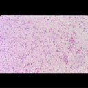
| 4. |
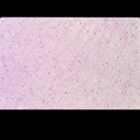
Representative photomicrograph from central portion of lesion
reveals relative acellularity. This corresponds to the low signal
intensity noted on the MR images. (Original magnification, X120
H-E stain) |
|
Copyright
©2003 All rights reserved.
The University of Texas M. D. Anderson Cancer Center
1515 Holcombe Blvd, Houston, TX 77030
1-800-392-1611 (USA) / 1-713-792-6161 Legal
Statements
Last updated; September
2003
|
|
|