|
|

|
|


|
| |
|

|
|
MIMICS
OF SOFT-TISSUE SARCOMAIN:
BENIGH LESIONS
|
|
INTRODUCTION
INTRAMUSCULAR
MYXOMA
- Benign, no
sex predilection
- Generally
occurs in age group > 40 years
- Thighs, shoulder,
gluteal region, arm
- Maybe associated
with fibrous dysplasia of the skeleton
- Prognosis
excellent, local recurrence rare
|
Click
on image(s) to view JPEG image(s).
|
| 1. |
Patient
8
47-year-old female with surgically proven intramuscular myxoma
of right proximal thigh.
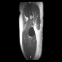
Sagittal T1W MR image reveals extremely low signal intensity
mass which is well defined. This extreme low signal intensity
on T1W MR images would be unusual for sarcoma and is a feature
of intramuscular myxoma. Differential diagnosis would include
rare intramuscular ganglion or cysts.
|
| 2. |
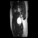
Sagittal T2W MR image reveals high signal intensity mass.
The high signal intensity is nonspecific unlike the extreme
low signal intenstiy noted on T1W MR images.
|
| 1. |
Patient
9
49-year-old female with surgically proven intramuscular myxoma
of leftl thigh.
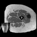
T1W
MR image reveals extremely low signal intensity well circumscribed
mass.
|
| 2. |
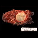
Gross
photograph of resected specimen. At surgery the intramuscular
myxoma contained gelatinous material.
|
|
Copyright
©2003 All rights reserved.
The University of Texas M. D. Anderson Cancer Center
1515 Holcombe Blvd, Houston, TX 77030
1-800-392-1611 (USA) / 1-713-792-6161 Legal
Statements
Last updated; September
2003
|
|
|