|
|

|
|


|
|
|
|

|
|
MIMICS
OF SOFT-TISSUE SARCOMA:
PSEUDOTUMORS
|
|
INTRODUCTION
NODULAR FASCIITIS
- Benign proliferation
of fibroblasts
- Most common
lesion of fibrous tissue
- Generally
presents as a rapidly growing solitary mass
- Most common
in age group 20-35 years
- Males = Females
- Occurs in
upper extremity (forearm), chest wall, back
- Head and
neck site more common in children
- Generally
< 3 cm in size, rarely larger size
- Occurs in
subcutaneous, intramuscular, fascial locations
- Treated by
local excision, spontaneous regression possible
|
Click
on image(s) to view JPEG image(s).
|
| |
Patient
1
39-year-old male with surgically proven nodular fasciitis
of right upper arm.
|
| 1. |
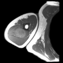
T1W
MR image reveals heterogenous intermediate signal intensity
mass (arrow on JPEG image) in posterior right upper arm.
|
| 2. |
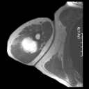
T2W
MR image reveals high signal intensity mass mimicking sarcoma.
MRI findings in nodular faciitis are nonspecific and biopsy
is necessary to establish diagnosis. Proliferative myositis
or fasciitis may also present as masses.
|
| 3. |
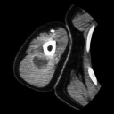
Contrast enhanced CT scan reveals mass with central low attenuation.
Low attenuation on CT scan in nodular fasciitis has been attributed
to myxomatous tissue component. |
| 4. |
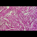
Representative photomicrograph from cellular portion of tumor
reveals haphazardly arranged proliferation of spindle shaped
fibroblastic cells. (Original magnification, X160 H-E stain)
|
| 5. |
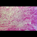
Representative photomicrograph from central portion of tumor
reveals loose connective tissue with a myxoid background. The
histological characteristics grossly correspond to the signal
intensity changes noted on the MR images. (Original magnification,
X120 H-E stain). |
| 6. |
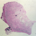
Photograph of histological cut section reveals cellular periphery
and cystic central region (arrow on JPEG image). |
|
Copyright
©2003 All rights reserved.
The University of Texas M. D. Anderson Cancer Center
1515 Holcombe Blvd, Houston, TX 77030
1-800-392-1611 (USA) / 1-713-792-6161 Legal
Statements
Last updated; September
2003
|
|
|
![]()