|
|

|
|


|
| |
|

|
|
POST
CHEMOTHERAPY (PRE-SURGERY)
|
|
Patient
2
44-year-old male with malignant fibrous histiocytoma of
right thigh initially diagnosed by needle biopsy.
Click on image(s) to view JPEG image(s).
|
| 1. |
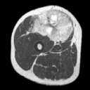
Axial T2W MR image reveals hyperintense chiefly solid tumor
with interspersed areas of 'necrosis' or cystic degenerative
change (arrows on JPEG image). |
| 2. |
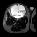
Axial T2W MR image 2 months post chemo/radiation therapy
reveals change in internal characteristics of tumor without
change in size secondary to therapy induced necrosis. The extensive
necrosis identified histologically precluded exact histological
characterization of tumor. |
|
Copyright
©2003 All rights reserved.
No part of this exhibit may be used for commercial purposes
without the expressed written consent of the Division of Diagnostic
Imaging, The University of Texas M. D. Anderson Cancer Center
1515 Holcombe Blvd, Houston, TX 77030
1-800-392-1611 (USA) / 1-713-792-6161 Legal
Statements
Last updated; September
2003
|
|
|