|
|

|
|


|
|
|
|

|
| POST-SURGICAL
APPEARANCE OF EXTREMITY SARCOMA |
|
INTRODUCTION
(continued)
If marginal
excision has been performed and an examination is performed prior
to subsequent wide excision, the goal of the radiologist is to adequately
assist in surgical planning. It is not necessary or possible to
attempt detection of minute foci of residual neoplasm but it is
essential to detect any significant residual tumor.
|
Patient
1
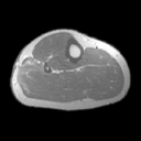
20-year-old female with MR study obtained following marginal
excision of parachordoma of leg.
Click on image to view JPEG image.
|
| 1. |

Axial T2W MR image reveals post surgical changes (arrow on
JPEG image) and no mass. At surgery following wide excision
minute microscopic residual tumor was identified. While MR imaging
is unable to detect such microscopic foci, gross foci may be
identifiable, thus aiding in surgical planning of wide excision. |
|
Copyright
©2003 All rights reserved.
The University of Texas M. D. Anderson Cancer Center
1515 Holcombe Blvd, Houston, TX 77030
1-800-392-1611 (USA) / 1-713-792-6161 Legal
Statements
Last updated; September
2003
|
|
|
![]()


