|
|

|
|


|
|
|
|

|
|
POST-
SURGICAL APPEARANCE OF EXTREMITY SARCOMA
|
|
Patient
5
40-year-old female with resected hemangiopericytoma of right
thigh.
Click on image(s) to view JPEG image(s).
|
| 1. |
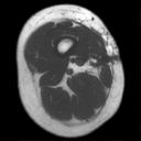
Axial T1W MR image obtained eight months post-surgery reveals
surgical scar (arrow on JPEG image) and noncontour deforming
post surgical changes within vastus medialis muscle in right
thigh. |
| 2. |
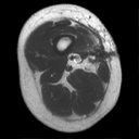
Post contrast T1W image reveals feathery enhancement in muscle.
Percutaneous aspiration performed two days following the MR
study did not reveal any recurrence. We do not know exactly
how much post-operative chemo/radiation therapy contributes
to the post therapy changes noted on MR imaging. |
|
Copyright
©2003 All rights reserved.
The University of Texas M. D. Anderson Cancer Center
1515 Holcombe Blvd, Houston, TX 77030
1-800-392-1611 (USA) / 1-713-792-6161 Legal
Statements
Last updated; September
2003
|
|
|