|
|

|
|


|
| |
|

|
|
GENERAL
IMAGING CHARACTERISTICS (PRE-THERAPY)
|
|
Patient
1
70-year-old female with surgically proven malignant fibrous
histiocytoma of right distal thigh and popliteal cyst.
Click on image(s) to view JPEG image(s).
|
| 1. |
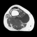
Axial T1W MR image reveals intermediate signal intensity
(relative to adjacent skeletal muscle) tumor (arrow on JPEG
image) and low signal intensity popliteal cyst (double arrows
on JPEG image). Most soft-tissue sarcomas exhibit intermediate
signal intensity on T1W MR images. |
| 2. |
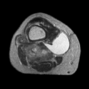
Axial T2W MR image reveals heterogeneous high signal intensity
sarcoma (arrow on JPEG image) and homogeneous considerably hyperintense
fluid containing cyst (double arrows on JPEG image). |
|
Copyright
©2003 All rights reserved.
No part of this exhibit may be used for commercial purposes
without the expressed written consent of the Division of Diagnostic
Imaging, The University of Texas M. D. Anderson Cancer Center
1515 Holcombe Blvd, Houston, TX 77030
1-800-392-1611 (USA) / 1-713-792-6161 Legal
Statements
Last updated; September
2003
|
|
|