|
|

|
|


|
| |
|

|
|
GENERAL
IMAGING CHARACTERISTICS (PRE-THERAPY)
|
|
Patient
3
53-year-old male with extraskeletal myxoid chondrosarcoma
of right posterior thigh.
Click on image(s) to view JPEG image(s).
|
| 1. |
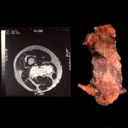
Axial T2W MR image (with surgically excised specimen) reveals
lobulated high signal intensity mass. While lobulation may be
seen in cartilaginous tumors or may be a feature of synovial
sarcomas, this finding is non-specific. |
| 2. |
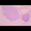
Representative low power photomicrograph from resected specimen
reveals two lobules of tumor (original magnification, X 60.
H-E stain). |
|
Copyright
©2003 All rights reserved.
No part of this exhibit may be used for commercial purposes
without the expressed written consent of the Division of Diagnostic
Imaging, The University of Texas M. D. Anderson Cancer Center
1515 Holcombe Blvd, Houston, TX 77030
1-800-392-1611 (USA) / 1-713-792-6161 Legal
Statements
Last updated; September
2003
|
|
|