|
|

|
|


|
| |
|

|
|
GENERAL
IMAGING CHARACTERISTICS (PRE-THERAPY)
|
|
Patient
4
71-year-old female with extraskeletal osteosarcoma of left
thigh.
Click on image(s) to view JPEG image(s).
|
| 1. |
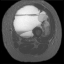
Axial T2W MR image reveals layering foci of hemorrhage within
tumor (arrow on JPEG image). Hemorrhage and cystic or 'necrotic'
changes in pre-treated sarcomas are non-specific features. |
|
Copyright
ę2003 All rights reserved.
No part of this exhibit may be used for commercial purposes
without the expressed written consent of the Division of Diagnostic
Imaging, The University of Texas M. D. Anderson Cancer Center
1515 Holcombe Blvd, Houston, TX 77030
1-800-392-1611 (USA) / 1-713-792-6161 Legal
Statements
Last updated; September
2003
|
|
|