|
|

|
|


|
| |
|

|
|
GENERAL
IMAGING CHARACTERISTICS (PRE-THERAPY)
|
|
Patient
8
45-year-old female with intramuscular lipoma (infiltrating
benign lipoma) of the proximal left thigh.
Click on image(s) to view JPEG image(s).
|
| 1. |
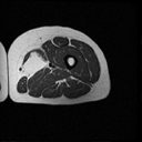
Axial T1W MR image reveals hyperintense fatty mass (arrow
on JPEG image) in left thigh. |
| 2. |
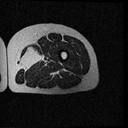
Axial T2W MR image reveals mass to be of similar signal
intensity as subcutaneous fat. Intramuscular lipoma may mimic
well differentiated liposarcoma. In our experience well differentiated
liposarcomas exhibit areas of high signal intensity on T2W images
and thickened septae within the lesion features not generally
noted in intramuscular lipomas. Treatment for both lesions is
the same and involves excision of the mass. |
|
Copyright
©2003 All rights reserved.
No part of this exhibit may be used for commercial purposes
without the expressed written consent of the Division of Diagnostic
Imaging, The University of Texas M. D. Anderson Cancer Center
1515 Holcombe Blvd, Houston, TX 77030
1-800-392-1611 (USA) / 1-713-792-6161 Legal
Statements
Last updated; September
2003
|
|
|