
|

|
REGIONAL
EXTENSION
Now viewing: Case
15
Case 15 - Recurrent squamous cell carcinoma of right orbit with
extension to ophthalmic division and gasserian ganglion
|
| This 68-year-old male had recurrent squamous cell
carcinoma in the right supra-orbital area. This was treated with local excision, orbital
exenteration, post-operative radiation therapy and chemotherapy. Recent follow-up MRI
examination showed progression of the orbital lesion with perineural extension to the
ophthalmic division of the trigeminal nerve and the gasserian ganglion. Patient was then
treated with continuation of selective chemotherapy. |
|
|
|
|
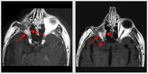
| Axial post-contrast T1-weighted MR image (TR 650/TE 16) at
the level of the mid-orbits: post operative status of the right orbit with
contrast-enhancing soft tissue tumor mass in the medial aspect of the orbit and
retrobulbar region (arrows). |
|
Axial post-contrast T1-weighted MR image (TR 650/TE 11) at
the level of the superior orbit fissure: contrast-enhancing soft tissue mass in the
retrobulbar region and superior orbit fissure (arrows). |
|
|
|
|
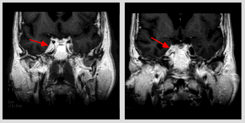
Coronal post-contrast T1-weighted MR image
(TR 500/TE 11) at the level of mid-sphenoid sinuses: contrast-enhancing soft tissue tumor
mass in the right cavernous sinus, mainly the region of V1 (arrow). |
|
Coronal post-contrast T1-weighted MR image (TR 500/TE 11) at
the level posterior to the sphenoid sinus: enlargement of the right gasserian ganglion due
to metastasis is well shown (arrow). |
|
|
©2002
The Levit Radiologic - Pathologic Institute
1100 Holcombe Blvd, Houston, TX 77030
(USA) / 713-792-2728
Last updated;
February 2002 - contact Webmaster
|
©2002
The University of Texas M. D. Anderson Cancer Center
1515 Holcombe Blvd, Houston, TX 77030
1-800-392-1611 (USA) / 1-713-792-6161 Legal
Statements
|
|
|
|