
|

|
REGIONAL
EXTENSION
Now viewing: Case
18
Case 18- Lymphoepithelioma of the nasopharynx with extensive
extension to maxillary and mandibular divisions of trigeminal and hypoglossal nerves
|
| Biopsy of the tumor mass of the nasopharynx
revealed lymphoepithelioma. Patient was treated with chemotherapy and followed with
radiation therapy. |
|
|
|
|
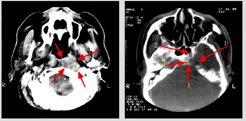
| Axial CT scan with IV contrast medium at the level of the
upper nasopharynx: there is a large soft tissue tumor involving the posterior and left
lateral walls of the nasopharynx with lateral and posterior extension (arrows). |
|
Axial CT scan with IV contrast medium at the level of the
base of the skull (bone window): there is extensive destruction of the clivus, foramen
lacerum, carotid canal, foramen ovale, and petrous pyramid on the left side (1). There is
also involvement of the posterior wall of the left sphenoid sinus (2). There is normal
appearance of the base of the skull on the right side. |
|
|
|
|
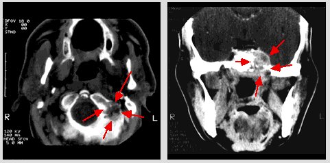
Axial CT scan with IV contrast medium at the
level of the foramen magnum: there is partial destruction of the left occipital condyle
including the hypoglossal canal (arrows). |
|
Coronal CT scan with IV contrast medium at the level of the
body of the sphenoid bone: tumor mass is extended superiorly to the left cavernous sinus
with involvement of the gasserian ganglion and laterally to the left foramen ovale
(arrows). The right foramen ovale and cavernous sinus are normal in appearance.
|
|
|
©2002
The Levit Radiologic - Pathologic Institute
1100 Holcombe Blvd, Houston, TX 77030
(USA) / 713-792-2728
Last updated;
February 2002 - contact Webmaster
|
©2002
The University of Texas M. D. Anderson Cancer Center
1515 Holcombe Blvd, Houston, TX 77030
1-800-392-1611 (USA) / 1-713-792-6161 Legal
Statements
|
|
|
|