|
|
|
|
|
|
||||||||||||||||
DISTANT
METASTASIS - The Trigeminal Nerve
|
||||||||||||||||

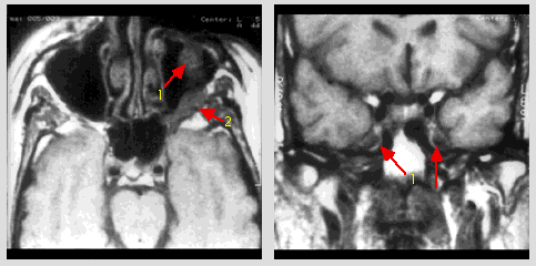
Case 2 - Recurrent squamous cell carcinoma of left cheek with metastasis to left maxillary division of trigeminal nerve |
|||||
| Operative findings: there was perineural metastasis to the maxillary division of the trigeminal nerve. The foramen rotundum was enlarged with a large maxillary division of the trigeminal nerve. The foramen ovale did not appear grossly abnormal, however, the mandibular division of the trigeminal nerve was somewhat thickened and enlarged. The gasserian ganglion was tight down upon the foramen ovale. A segment of the second division of the trigeminal nerve was removed for frozen section. Histopathology revealed infiltrative squamous cell carcinoma, neural invasion. | |||||
|
|||||
|
©2002
The Levit Radiologic - Pathologic Institute Last updated; February 2002 - contact Webmaster |
©2002
The University of Texas M. D. Anderson Cancer Center 1515 Holcombe Blvd, Houston, TX 77030 1-800-392-1611 (USA) / 1-713-792-6161 Legal Statements
|