Case 3 - Adenoid cystic carcinoma of left maxillary sinus with
metastasis to maxillary division and gasserian ganglion
Open biopsy of the left maxillary sinus revealed
adenoid cystic carcinoma. The case was deemed inoperable and treated with radiation
therapy and chemotherapy.
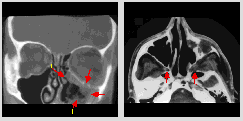
Coronal CT scan with IV contrast medium at the level of the maxillary and
mid ethmoid sinuses: there is postoperative change in the left maxillary sinus and nasal
fossa with absence of inferior turbinate. A large irregular soft tissue mass occupying
most part of the left maxillary sinus with destruction of the bony walls (1). The
left infraorbital canal is enlarged with destruction of the bony boundary (2).
Axial CT scan with IV contrast medium at the level of the
maxillary sinus and nasopharynx: soft tissue mass in the left pterygopalatine fossa with
destruction of the bony boundary (1). The right pterygopalatine fossa is normal (2).
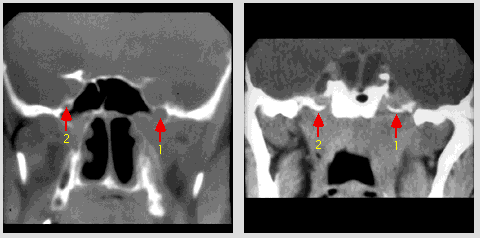
Coronal CT scan with IV contrast medium at the level of the anterior
sphenoid sinus: there is enlargement and erosion of the left foramen rotundum (1). The
right foramen rotundum is normal (2).
Coronal CT scan with IV contrast medium at the level
posterior to the dorsum sella: there is enlargement of the left gasserian ganglion (1).
The right ganglion is normal (2).