Scientific Exhibits
Home


Return to the top of
Section VII


View Previous Case

View Next Case


DISTANT METASTASIS - The Trigeminal Nerve

Now viewing: Case 4

Case 4 - Recurrent adenoid cystic carcinoma of left submandibular gland with metastasis to left mandibular division and gasserian ganglion

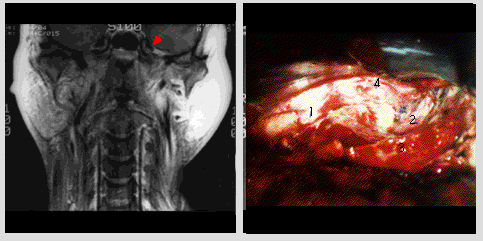
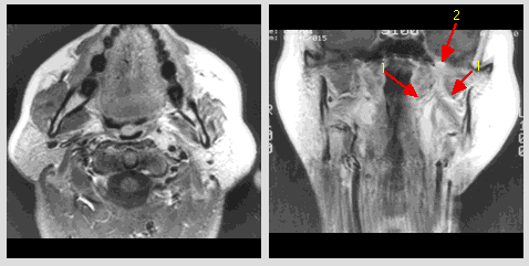
This 55-year-old male had a history of adenoid cystic carcinoma of the left submandibular gland which was resected and followed by post operative radiation therapy, 3 years ago. Because of persistent pain in the operated area extending to the temporal region and paresthesia along the left mandible follow-up CT and MRI of the head and neck were taken and showed tumor recurrence with perineural metastasis to the left mandibular division of the trigeminal nerve extending to the gasserian ganglion. Left frontotemporal craniectomy with extradural exploration of mandibular division of the trigeminal nerve and gasserian ganglion were performed. There was tumor mass involving the mandibular division of the trigeminal nerve and extending to the gasserian ganglion. Multiple microbiopsies were done in the mandibular division. Histopathology revealed perineural invasion by adenoid cystic carcinoma.
case_04.gif (27161 bytes)
Axial post-contrast T1-weighted MR image (TR 500/TE 16) at the level of the submandibular region: post radiation and post operative changes in the left submandibular region without evidence of recurrence. Coronal post-contrast T1-weighted MR image (TR 800/TE 11) with ChemSat fat suppression, at the level of the foramen ovale: enlargement of the branches of the mandibular division of the left trigeminal nerve with high signal intensity (1) and soft tissue densities with intermediate signal intensity at the left base of the skull in the region of the foramen ovale (2). There is widening of the left foramen ovale and enlargement of the mandibular division of the left trigeminal nerve with high signal intensity.
case_04a.gif (40944 bytes)
Coronal post-contrast T1-weighted MR image (TR 800/TE 11) ChemSat fat suppression at the level of the posterior aspect of the sphenoid sinuses:   enlargement of the left gasserian ganglion with high signal intensity is well shown (arrow). Photograph of the tumor mass of the left trigeminal nerve and gasserian ganglion:
1.Maxillary division of the trigeminal nerve at foramen rotundum
2.Mandibular division of the trigeminal nerve at foramen ovale
3.Area of biopsies
4.Gasserian ganglion


©2002 The Levit Radiologic - Pathologic Institute
1100 Holcombe Blvd, Houston, TX 77030
 (USA) / 713-792-2728     

Last updated; February 2002 - contact  Webmaster 

©2002 The University of Texas M. D. Anderson Cancer Center
1515 Holcombe Blvd, Houston, TX 77030
1-800-392-1611 (USA) / 1-713-792-6161     Legal Statements