![]()
![]()
| Introduction | Section 1 | Section 2 | Section 3 | Conclusion and References |
|
View Table
of contents
|
Section 1 - Direct
leukemic involvement of the Central Nervous System Nonmeningeal disease: Brain parenchyma |
|
|
Click on image to enlarge
|
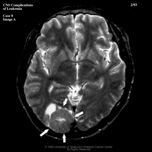
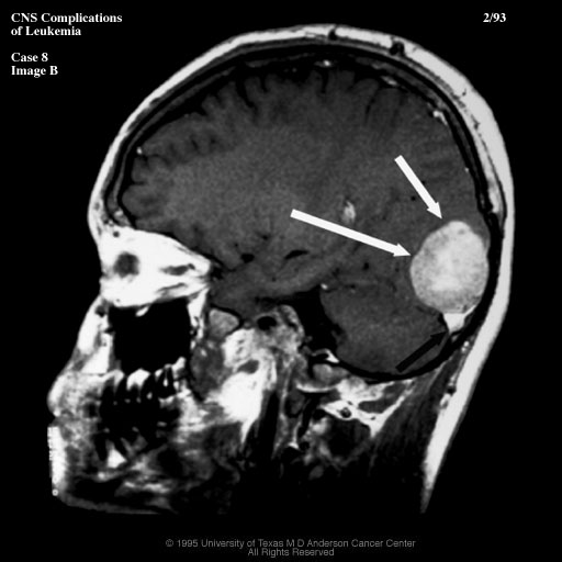
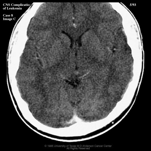
Case 8 This patient initially presented with fatigue, blurred vision and headache. BM aspiration revealed blast cells, and the dx of AML was made. MRI (8A:T2, 8B:T1 post) showed a large mass (white arrows), probably extraaxial, in the right occ. region which was isointense on T1 and T2, and enhanced brightly, mimicking a meningioma.
Note the dural sinus thrombosis.This lesion resolved completely with chemotherapy (8C:CECT), and was almost certainly a chloroma. |
|
|
©2002
The Levit Radiologic - Pathologic Institute Last updated; February 2002 - contact Webmaster |
©2002
The University of Texas M. D. Anderson Cancer Center
|
|