|
New Multiprobe FISH Test
on Urologic Specimens is Highly Sensitive in the Diagnosis of Urothelial
Carcinoma *
Abha Khanna, Aazam Alizadeh, Jun Gu, Feng Jiang, Hua-Zhong
Zhang, Arnout Ruifrok, Nancy Caraway, Dennis Johnston, Ruth L Katz. University
of Texas, M.D.Anderson Cancer Center, Houston, TX; University of Texas,
M. D.Anderson Cancer Center, Houston, TX.
Introduction
Urologic cytology is not sensitive in diagnosing urothelial
carcinoma (UC). Fluorescence-in-situ-hybridization (FISH) for multiple
centromeric probes has previously been shown to be a very sensitive test
for diagnosing UC, however the test was limited by the requirement of multiple
cytospins to evaluate 4 or more probe sets. Recently a new commercial
test (VYSIS) for evaluating urinary cytology became available in which
4 probes are simultaneously evaluated on a per cell basis on a single cytospin.
We performed a pilot study to test the efficacy of the new FISH test compared
to standard urine cytology.
Materials and Methods
14 urinary specimens were reacted with a DNA probe set
labeled with different fluorochromes and directed against the chromosomes
3,7, and 17, and a locus specific probe against 9p21. 25 cells were
scored for each probe. Normal control bladder wash showed 2 signals
(diploid) for each probe. Abnormal signals were defined as non-diploid.
Results.
11/14 specimens were evaluable. The number of abnormal
cells ranged from 14 to 25 with a median of 22 abnormal cells. Of
6 cases with 22 or less abnormal cells, 4 were scored as atypical by cytology
and 2 as UC. All 5 cases with >2 abnormal cells were high grade UC.
Two cases of high grade UC showed 90% homozygous deletion for 9p21, while
the remaining cases showed a wide range of LOH for 9p21.
Conclusion
This study showed that the multi-color FISH probe test
was more sensitive than cytology, easily performed and yielded a high number
of cells with numerical chromosomal aberrations. Further studies
are needed to evaluate these results prospectively against follow-up biopsies.
*This abstract has been selected for platform presentation
at the 48th Annual Scientific Meeting of the American Society of Cytopathology,
Philadelphia,Nov.7-11, 2000.
Reference: The Development of a Multitarget, Multicolor
Fluorescence in Situ Hybridization Assay for the Detection of Urothelial
Carcinoma in Urine. Irina A et.al. Journal of Molecular Diagnostics, (2)3
116-123, 2000)
 |
 |
 |
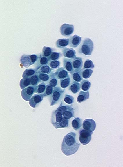
| Figure 1. Cytology from a normal urine specimen showing
normal morphology. |
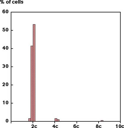
Figure 2. Corresponding diploid DNA histogram from the
same specimen shown left . |
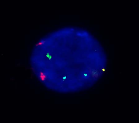
Figure 3. Normal UroVysion FISH pattern from the same
specimen shown left. |
 |
 |
 |
| Figure 4. Cytology from a low grade UC patient showing
apparently normal morphology. |
Figure 5. Corresponding false negative diploid DNA histogram
from the same specimen shown left. |
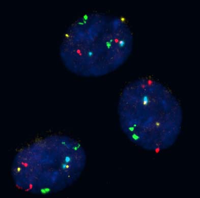
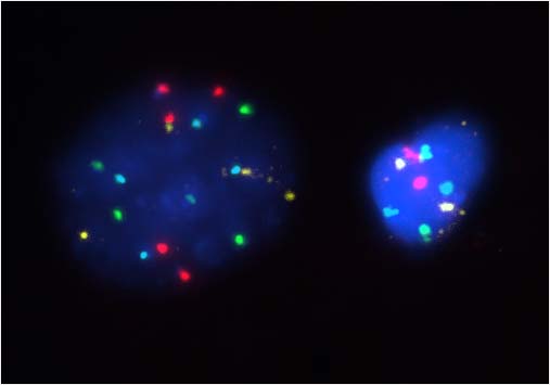
Figure 6. Aaneuploid UroVysion FISH pattern from the
same specimen shown left. |
 |
 |
 |
| Figure 7. Cytology from a patient with high grade UC
showing abnormal morphology. |
Figure 8. Corresponding aneuploid DNA histogram from
the same patient shown left . |
Figure 9. Aaneuploid UroVysion FISH pattern from the
same patient shown left. |
How to request the FISH test
When urothelial carcinoma is suspected, you may request this
highly sensitive FISH test by calling Research Image Diagnostic Lab at
(713)792-4087 or (713) 792-4088. Or you can page us at (713)404-7095 and
someone will return your call. We prefer to receive 150 ml voided urine
or bladder washes mixed with same amount of 50% ethanol. We will provide
specimen collection kits for outside requester.
|