![]()
![]()
| Introduction | Section 1 | Section 2 | Section 3 | Conclusion and References |
|
View Table
of contents
|
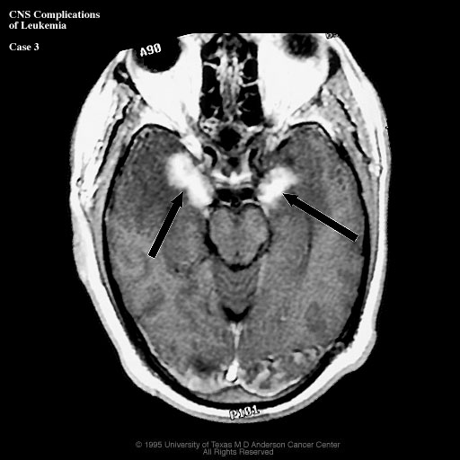
Section 1 - Direct
leukemic involvement of the Central Nervous System Meningeal disease: Leukemic infiltration of the meninges |
|
|
Click on image to enlarge
|
Case 3
|
|
|
©2002
The Levit Radiologic - Pathologic Institute Last updated; February 2002 - contact Webmaster |
©2002
The University of Texas M. D. Anderson Cancer Center
|
|