![]()
![]()
| Introduction | Section 1 | Section 2 | Section 3 | Conclusion and References |
|
View Table
of contents
|
Section 1 - Direct
leukemic involvement of the Central Nervous System |
|
Meningeal disease: Leukemic infiltration of the meninges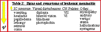 Diagnosis:The diagnosis of leukemic meningitis generally depends on the detection of leukemic cells in the CSF, however cytology can be falsely negative. Imaging may be necessary. Findings include hydrocephalus (may be the only finding), or an abnormal MR appearance of CSF on precontrast imaging. If CSF is not of appropriate signal intensity on T1 and T2-weighted images, the possibility of leukemic meningitis should be raised. Abnormal meningeal enhancement, in the cisterns or along the pial surface of the brain or spinal cord, is the surest sign of leukemic meningitis (cases 1-3). This may be smooth or nodular. MRI is known to be far more sensitive in the detection of leptomeningeal tumor spread than CT, however CT can make this diagnosis in flagrant cases. Be aware that this type of enhancement is nonspecific (see pitfalls). Dural spread of leukemia is best detected radiologically as abnormally thickened and brightly enhancing dura on Gd-enhanced MRI.
|
||
|
©2002
The Levit Radiologic - Pathologic Institute Last updated; February 2002 - contact Webmaster
|
©2002
The University of Texas M. D. Anderson Cancer Center
|
|