Detection of Cyclin D1 Translocation
by FISH is Highly Specific and Sensitive in the Diagnosis of Mantle Cell
Lymphoma (MCL)
Ruth L. Katz, M.D., Director
Image Cytometry/FISH Diagnostic Laboratory, University
of Texas, M.D. Anderson Cancer Center
Background
-
Mantle cell lymphoma (MCL) is a subtype of non-Hodgkins
lymphoma characterized by poor prognosis and a median survival of approximately
3 years.
-
On the basis of morphology and immunophenotype alone, mantle
cell lymphoma is difficult to distinguish from indolent lymphomas and leukemias.
-
Cytogenetically, a t(11:14)(q13;q32) is associated with 75%
of mantle cell lymphomas.
-
PCR for the major translocation cluster (MTC) region of the
bcl-1
is positive in only 30-40% of cases.
-
Southern blot analysis detects translocations in approximately
50% to 70% of the cases.
-
Translocation breakpoints are scattered within the approximately
120-kb bcl-1 region adjacent to cyclin D1, whereas part of
them are clustered in the MTC.
-
The translocation leads to overexpression of cyclin
D1 due to juxtaposition of the Ig heavy chain gene enhancer on 14q32 to
the cyclin D1 gene on 11q13.
Cyclin D1/CEP11
-
Cyclin D1 amplification is common in many types of
cancer, second only to the HER2/neu (erb-b2) oncogene.
-
The LSI Cyclin D1 is approximately 300 kb in size.
-
This probe may be used to determine the copy number of the
Cyclin
D1 locus, or as an enumerator probe for chromosome 11 in interphase and
metaphase cells.
-
200 cells will be counted. The result will be expressed as
the ratio of Cyclin D1/CEP11.
Cyclin D1FISH test report
format
Print Date: 06/04/1999
Name: ##############
Location: DICT
Hospital Number: (00000)#########
Attending : #############
Age: 50 YRS Sex: F
CYTOPATHOLOGY - PHONE: (713)794-5625
LYMPHOPROLIFERATIVE DISORDER FISH ANALYSIS
ACCESSION # : FI-99-0060
COLLECTION DATE: 5/27/1999
RESULTS:
SAMPLE TYPE: Bone marrow
Signals
Probe
1 2
3 4
5 6
7 8
>8
11CEP(%)
1 98
1 0
0 0
0 0
0
Cyclin D1(%)
0 13.5 87
0 0
0 0
0 0
200 cells counted
For Cyclin D1 test:
Ratio = Total Cyclin D1/Total 11CEP = 287/200
= 1.43
Normal range: Mean ratio +/- SD = 0.99 +/- 0.09.
95% Confident Interval: (0.96, 1.02)
Ratio for Mantle cell positive control: 1.49
Ratio for Mantle cell negative control: 1.00
ISCN DIAGNOSIS: nuc ish 11cen(D11Z1 x 2), 11q13(CCND1
x 3) [200]
INTERPRETATION:
Elevated ratio Cyclin D1: total 11 CEP at 1.43 with
87% cells showing evidence of Cyclin D1 translocation consistent with extensive
involvement by mantle cell lymphoma in bone marrow.
Notes: Based on mixing experiments, in peripheral blood,
bone marrow, or lymph-node specimens, the level of detectable translocation
t(11;14)(q13;q32) cells is considered positive if 5% or higher numbers
of translocated cells are presented.
The in situ hybridization (ish) technique was performed
using a Vysis LSI Cyclin D1/CEP11 probe. A total of 200 interphases were
analyzed.
In situ hybridization is for investigational use only
and should not be used for diagnostic purposes without confirmation by
another proven procedure. This test does not rule out abnormalities other
than the one for which the probe was designed.
REFERENCE: R.L.Katz, J.Gu, L.Pasco-Miller, H.Z.Zhang,
N.Caraway, and L.J.Medeiros. Detection of Cyclin D1 Translocation by Fluorescence
in-situ Hybridization (FISH) is Highly Specific and Sensitive in the Diagnosis
of Mantle Cell Lymphoma (MCL) Modern Pathology Vol.12.1999.
Date: Verified by:
CAC (Electronic Signature)
* Result will be reported on the Cerner in about a week after sample received.
Fluorescence
in
situ Hybridization (FISH) Analysis
 |
 |
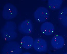
| Figure 1. Interphase FISH from a normal cell line. Cells
showing 2 green signals (Centromere 11) and 2 orange signals (Cyclin D1
at chromosome 11q13). |
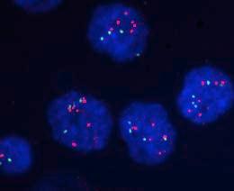
Figure 2. Interphase FISH from a non-MCL patient. Cells
showing 2 green signals (Centromere 11) and 2 orange signals (Cyclin D1
at chromosome 11q13). |
 |
 |
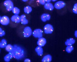
| Figure 3. Another interphase FISH from a non-MCL patient.
Cells showing 2 green signals (Centromere 11) and 2 orange signals (Cyclin
D1 at chromosome 11q13). |
Figure 4. Interphase FISH from a cell line with cytogenetically
detectable t(11;14)(q13;q32). Most of cells showing 2 green signals (Centromere
11) and 3 orange signals (Cyclin D1 at chromosome 11q13). |
 |
 |
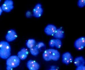
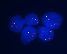
| Figure 5. Interphase FISH from a MCL patient with tetraploid
cell population. Most of the cells showing 4 green signals (Centromere
11) and 5 orange signals (Cyclin D1 at chromosome 11q13). |
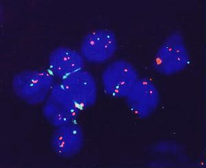
Figure 6. Interphase FISH from a blastoid MCL patient.
Cells are showing 2 green signals (Centromere 11) and 3 or more orange
signals (Cyclin D1 at chromosome 11q13). |
How to request the FISH test
When Mantle Cell Lymphoma is in the differential diagnosis,
you may request this highly sensitive FISH test by calling Research Image
Diagnostic Lab at (713)792-4087 or (713) 792-4088. Fine needle aspiration
samples in culture media or PBS, peripheral blood samples in sodium heparin
vacutainer, or bone marrow samples in sodium heparin vacutainer will be
acceptable for the test. |