|
Comparison of two Fluorescence-In-Situ-Hybridization
(FISH) approaches in the detection of t(11;14)(q13;q32) for Mantle Cell
Lymphoma (MCL) *
Jun Gu, Tanweer M Zaidi, Huihui Shi, Abha Khanna,
Jorge Romaguera, Nancy P Caraway, Feng Jiang, Hua_Zhong Zhang, Issa F Khouri,
Ruth L Katz. University of Texas, M.D.Anderson Cancer Center, Houston,
TX; University of Texas, M. D.Anderson Cancer Center, Houston, TX.
Segregation and fusion interphase FISH are two frequently
used methods to detect chromosomal abnormalities for lymphomas and leukemia.
Previously we demonstrated that Cyclin D1/CEP11 ratio FISH (rFISH) is highly
sensitive in the diagnosis of MCL. However, this is an indirect approach
and has shown about 10% false negative results based on corresponding flow-cytometry
(FCM) and histology due to the fact that t(11;14)(q13;q32) may occur without
splitting of cyclin D1. Fusion FISH (fFISH) by directly detecting the t(11;14)(q13;q32)
fusion signal, avoids the false negative rate by rFISH. In this study we
performed paired rFISH and fFISH on 15 peripheral blood (PB) and 24 bone
marrow (BM) samples from patients with confirmed MCL. At least one fusion
signal >14% was determined as a cut off by testing PB lymphocytes from
40 normal individuals. Of 39 samples tested, 23 (59%) were fFISH+. However
of these 23, only 12 (52%) were rFISH+. Of 11 samples with confirmed positive
histology and FCM, 10 (91%) were fFISH+, 7 (64%) were rFISH+. The percentage
of cells with fusion signal for fFISH ranged from 2% to 82.5% with predominantly
single fusion signal. The percentage of double fusion signals increased
dramatically only when rFISH ratio > = 1.10. Our study showed that fFISH
is more sensitive than rFISH and therefore is better for the detection
of minimum residue disease (MRD). Although double fusion, an indication
of reciprocal t(11;14)(q13;q32) translocation is more specific, single
fusion is as important as double fusion in the diagnosis of MCL. The use
of fFISH will significantly increase our power to detect MRD with t(11;14)(q13;q32)
in patients with MCL.
*This abstract has been submitted to 92nd Annual Meeting
of American Association for Cancer Research, New Orleans,March 24-28,2001
Fluorescence
in
situ Hybridization (FISH) Analysis
 |
 |
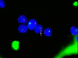
| Figure 1. Interphase fFISH from MCL patient with negative
rFISH result showing fusion signal between CCND1 (11q13, orange) and IgH(14q32,
green). |
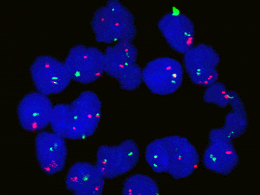
Figure 2. Interphase fFISH from a MCL patient with positive
rFISH result showing either single and double fusion between CCND1 (11q13,
orange) and IgH(14q32, green). |
How to request the FISH test
When Mantle Cell Lymphoma is in the differential diagnosis,
you may request this highly sensitive FISH test by calling Research Image
Diagnostic Lab at (713)792-4087 or (713) 792-4088. Fine needle aspiration
samples in culture media or PBS, peripheral blood samples in sodium heparin
vacutainer, or bone marrow samples in sodium heparin vacutainer will be
acceptable for the test. |